
 首页
首页 登录
登录 注册
注册


 下载:
下载:
-
压力作为热力学的重要基本参数之一[1],几乎对材料的化学、结构、机械、电子、磁性、声子等所有属性[2-3]均可以产生影响,因此在物理[4]、化学[5]、材料科学[6]及地球科学[7]等多个研究领域都得到了广泛应用。利用金刚石对顶砧压腔(Diamond anvil cell,DAC)产生的高压研究样品的性质是实验室最常用的高压研究手段之一。在金刚石压砧的作用下,DAC样品腔内可以达到几十万甚至几百万大气压的极高压力[8-9],压腔内的微量样品能够发生多次物性变化。然而,受DAC的结构及样品材料等的限制,高压研究必须在窗口有限、样品尺度微小且X射线能够穿过环境材料(如金刚石压砧、传压介质)等条件下进行。这些限制使得具有能量范围宽、通量高、准直性好、能量带宽可调、有时间结构及相干性好等特性的同步辐射装置在高压研究中发挥了重要作用。
自20世纪80年代以来,同步辐射技术与DAC技术相结合,为探索高压条件下的材料物性提供了丰富的研究手段[2-3,10]。例如:高压X射线衍射(X-ray diffraction,XRD)方法可用于确定晶体或非晶材料的结构、密度、应力应变和择优取向等信息[11];X射线吸收谱学(X-ray absorption spectroscopy,XAS)可以针对材料中特定元素进行结构表征,获得感兴趣元素的局域结构信息(价态、构型、配位数、键长及无序度等)[12-13];X射线核共振散射谱学(Nuclear resonance scattering,NRS)可提供时域的Mössbauer超精细结构谱学信息以及声子投影态密度[14];X射线拉曼散射(X-ray raman scattering,XRS)可探测轻元素(C、N和O等)在高压条件下的化学键变化[15];X射线成像(X-ray imaging,XRI)可以研究样品在高压条件下的物态方程、相演变及其动力学过程[16]。
同步辐射光源技术的不断发展,也在推动高压科学的不断进步。近年来,国际主要高能光源,如美国先进光子源(APS)[17]、欧洲同步辐射光源(ESRF)[18]、日本的Spring-8光源[19]和德国的PETRA Ⅲ光源[20],都在积极实施或推进升级具有准衍射极限环的第四代同步辐射光源计划。高能同步辐射光源(High energy photon source,HEPS)[21-22]作为我国“十三五”期间建设的、为国家重大战略需求和前沿基础科学研究提供技术支撑平台的国家重大科技基础设施,于2017年12月获得国家发展改革委批复立项,并于2019年6月在北京怀柔奠基启动建设,计划将于2025年完成建设并投入使用。作为第四代同步辐射光源,HEPS光源具有极小的发射度,能够提供比现有第三代同步辐射光源亮度高100倍以上的同步辐射光,实验站也更容易获得微米和亚微米(纳米)尺度的聚焦光斑。同时,低发射度光源具有的相干性优势也将极大地促进相干谱学、相干成像等实验技术的发展。这些优异的性能可以为高压科学在更高压力范围、更小时间或空间尺度等条件下开展研究提供重要支撑,例如:极高压(太帕量级)条件下的物性研究、压力(或温度)快速加载条件下的时间分辨研究、极高压条件下的局域变化及不均匀性研究、地球(行星)深部温压条件下的物质研究等[3]。
高压科学研究将是HEPS建成后的一个重要应用方向。本文的主要目的是向高压领域相关科研工作者介绍HEPS一期建设过程中与DAC高压实验技术相关的线站设计。一方面,有助于用户更多地了解HEPS,为将来在HEPS上开展高压研究工作做一些初步的准备;另一方面,也希望得到用户对目前线站设计方案的反馈,包括对未来二期、三期线站布局中高压光束线站规划的意见与建议。
全文HTML
-
HEPS是具有极低发射度的第四代高能同步辐射光源,于2019年6月29日在北京市怀柔区动工,并计划于2025年底完成验收。HEPS的设计亮度大1022 phs·s–1·mm–2·mrad–2·(0.1%B.W.)–1,自然发射度为34.2 pmrad,具体参数见表1。HEPS建成后能够对微观结构从静态构成到动态演化,提供多维度、实时、原位表征,解析物质结构生成及演化的全周期全过程,揭示微观物质结构的生成演化机制,剖析微观物质构成,为物质调控提供基础,从而推动材料科学、化学工程、能源环境、生物医学等领域的科学研究。
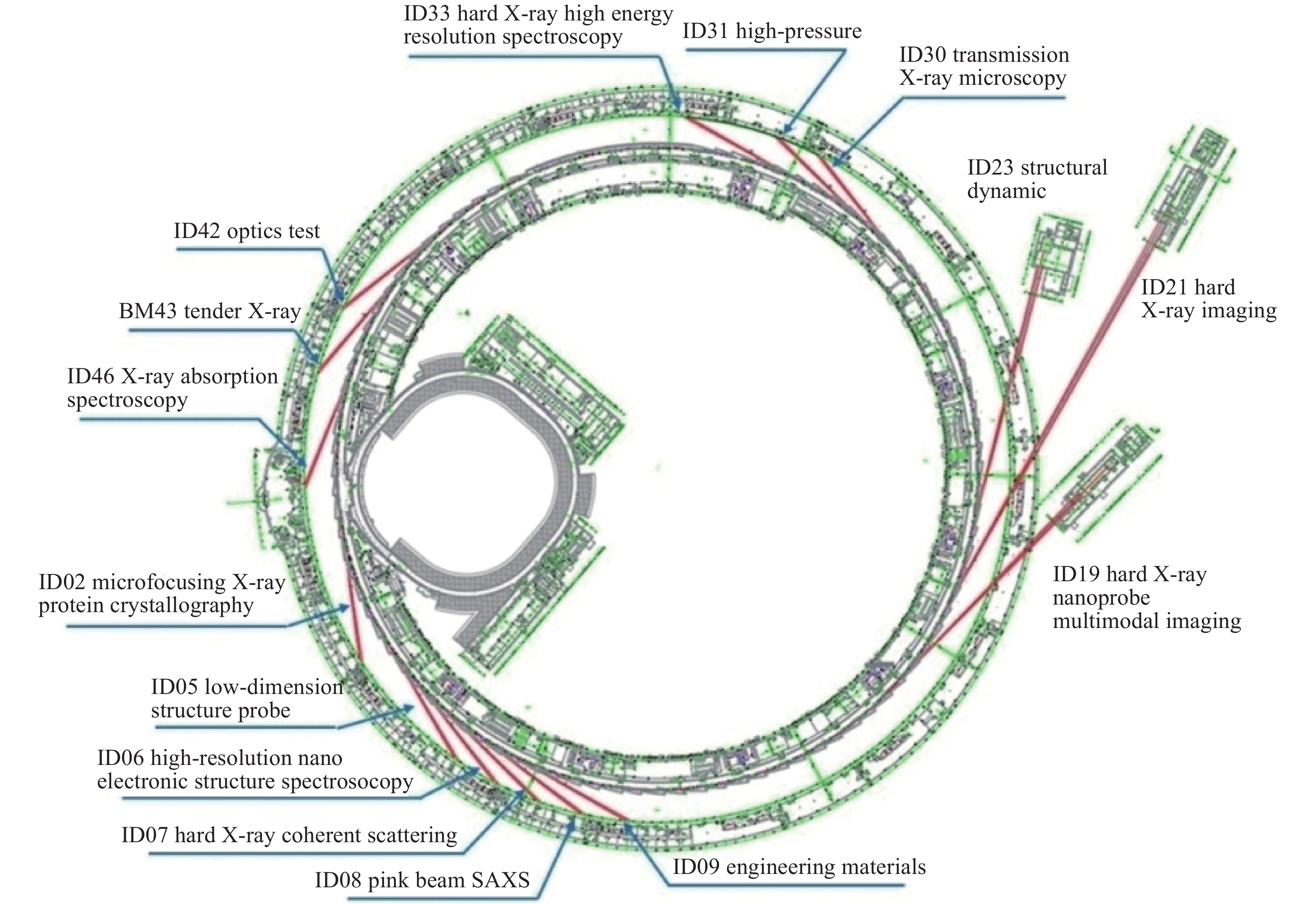
HEPS建成后将具备90条以上高性能光束线站的容量,其中一期建设的线站共14条(另外包括一条测试束线)。图1所示为HEPS一期线站布局,其中以ID开头的线站是在直线节安装插入件的引出线站,以BM开头的线站为弯铁引出线站。3条延伸到实验大厅以外的长光束线能在纳米聚焦、相干、时间分辨到高能等方面充分发挥新光源优势。HEPS一期线站的规划主要是在考虑体现新光源高能、高亮度等优势,满足国内用户群体需求及“衍射极限光源先进的实验方法在实验站全覆盖”等原则基础上完成的。
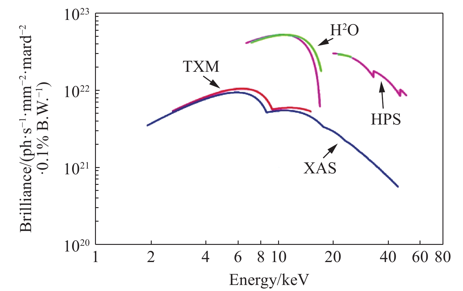
限于篇幅,本文将重点介绍HEPS一期建设线站中与高压学科最为相关的若干线站,分别是:X射线显微成像线站(ID30)、高压线站(ID31)、硬X射线高分辨谱学线站(ID33)和X射线吸收谱学线站(ID46)。其中,高压线站是高压X射线衍射专用实验站,其他线站可以通过优化设计兼容高压实验方法。图2所示为这些线站建成后插入件的“能量-亮度”谱,其中:HPS表示高压线站,H2O表示硬X射线高分辨谱学线站,XAS表示X射线吸收谱学线站,TXM表示X射线显微成像线站。
-
高压光束线站(High-pressure beamline,HPB)从HEPS储存环插入件ID31引出,是开展以高压XRD实验为主的专用高压线站。实验站将以XRD方法为基础建立多种高压研究手段,包括粉末XRD、单晶XRD、径向XRD、时间分辨以及对分布函数(Pair distribution function,PDF)测量等方法,同时还将结合激光加热、电阻加热、低温、压力动态加载等样品环境,建立多种原位条件的XRD实验方法。线站建成后,将与国际上主要高能同步辐射光源升级后的高压线站(如APS的GSECARS[23]和HPCAT[24]线站、ESRF的ID27[25]线站、Spring-8的BL10XU[26]线站和PETRA Ⅲ的P02.2[27]线站等)具备同等水平的实验及数据分析条件。
在应用同步辐射技术的高压科学研究中,XRD是最基础、最常用的实验方法之一。单晶、多晶/粉晶、纳米晶体以及非晶体或液体都可以成为高压XRD的实验样品,它能够准确提供样品在高压条件下的结构信息。利用高压XRD方法,也能够对样品的状态方程、晶体结构、相转变、弹性和晶格应变等开展研究。利用第四代衍射极限同步辐射光源提供的小尺寸(小于1 μm)、高通量X射线光斑,利用高压XRD方法还可以开展超高压(大于500 GPa)、局域结构变化、压力或温度动态加载条件下的结构变化等研究工作。
-
HPB建成后可以为用户提供20、30、40和50 keV 5种能量点的X射线光源,X射线聚焦光斑尺寸为十几微米到亚微米(约150 nm),样品处的光子数大于1013 ph/s。根据用户需要,也可以提供大尺寸(约200 μm)的聚焦光斑。实验站还可以实现4~4 000 K温度范围的原位高压XRD实验。
结合压力动态加/卸载以及脉冲激光加热技术,实验站还可以为用户提供曝光小于10微秒每帧的动态实验条件。在压力动态加载实验中,压力的加载速率可以达到150 TPa/s以上。
-
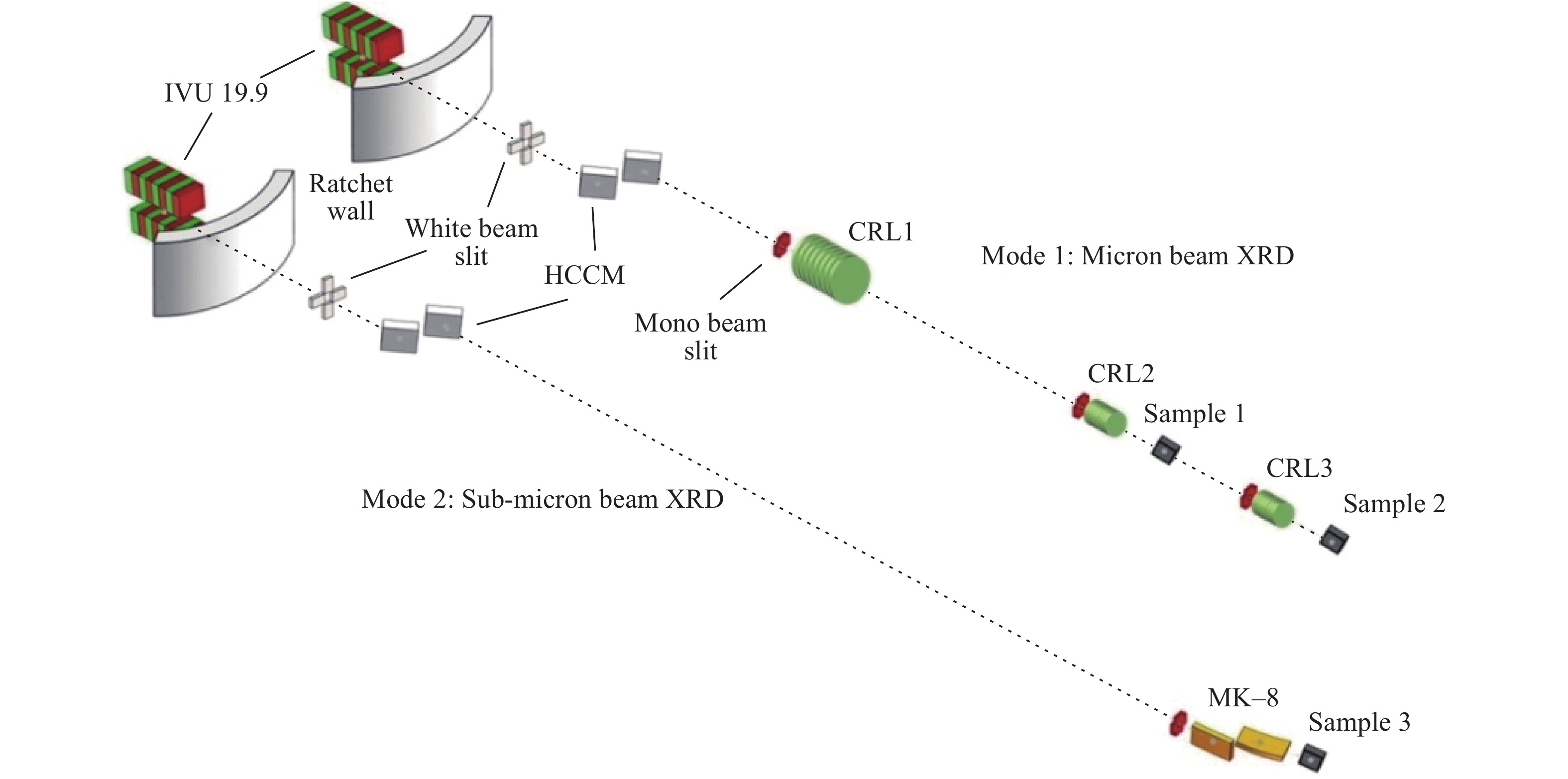
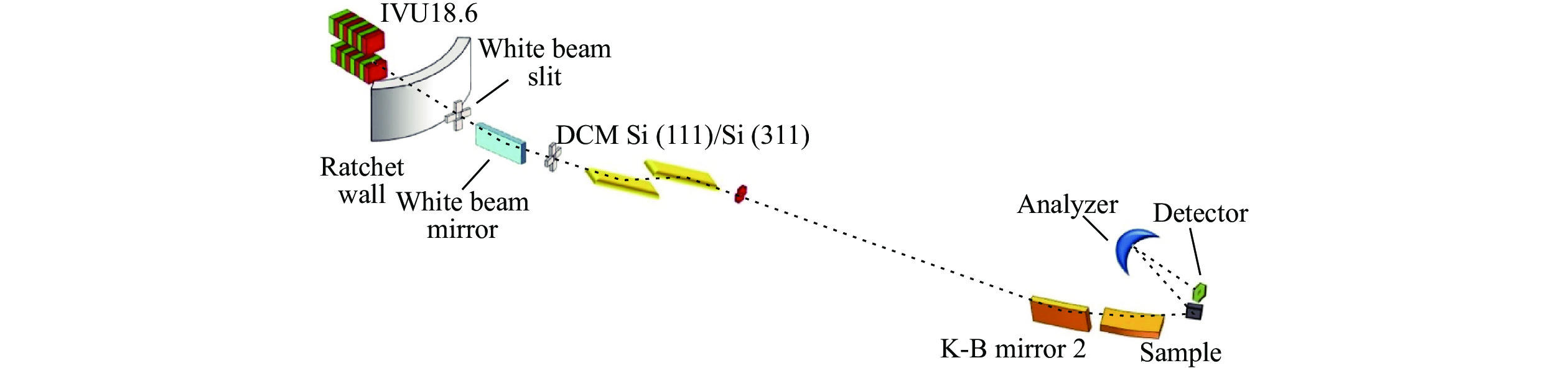
HPB光学布局如图3所示,X射线从储存环的低β直线节ID31引出。真空内波荡器(IVU)的磁周期长度为19.9 mm,总周期数201,插入件总长度4 m,设计最小磁场间隙5.2 mm。经过单色器选取指定能量的单色光后,由聚焦元件聚焦到样品位置。水平反射的channel-cut单色器(HCCM)采用液氮冷却,不仅能承受高热负荷,还可以提供高稳定性的单色光输出,对提高样品点位置X射线聚焦光斑的稳定性有很大帮助。通过两种不同聚焦元件获得X射线聚焦光斑:利用复合折射透镜(Compound refractive lens,CRL)获取微米聚焦光斑,利用多层膜K-B聚焦镜(Multilayer K-B mirrors,MK-B)获取亚微米聚焦光斑。
高压光束线站有两个微米聚焦样品点,分别位于距光源点84 m的Sample 1及距光源点89 m的Sample 2。通过两级CRL对X射线顺序聚焦获得微米光斑,CRL1+CRL2完成Sample 1处X射线聚焦,CRL1+CRL3完成Sample 2处的聚焦。Sample 1和Sample 2的光斑半高宽一般为2 μm左右,如果需要更大的光斑,可以通过移动CRL2和CRL3的位置实现。亚微米聚焦光斑位于距光源点95 m的Sample 3处,可由MK-B直接聚焦获得。亚微米聚焦光斑的半高宽约为150 nm,从聚焦镜后端到样品的工作距离大于100 mm,可以满足大多数原位高压XRD实验的需求。
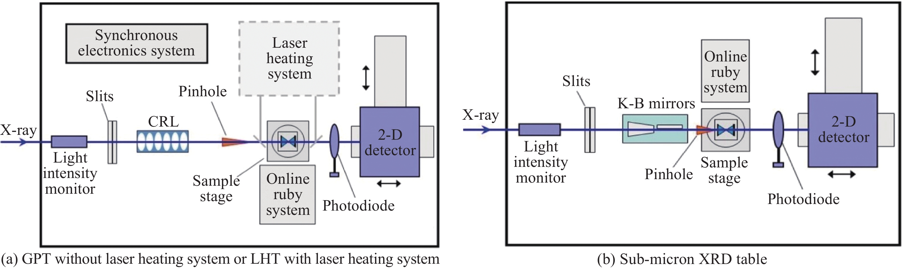
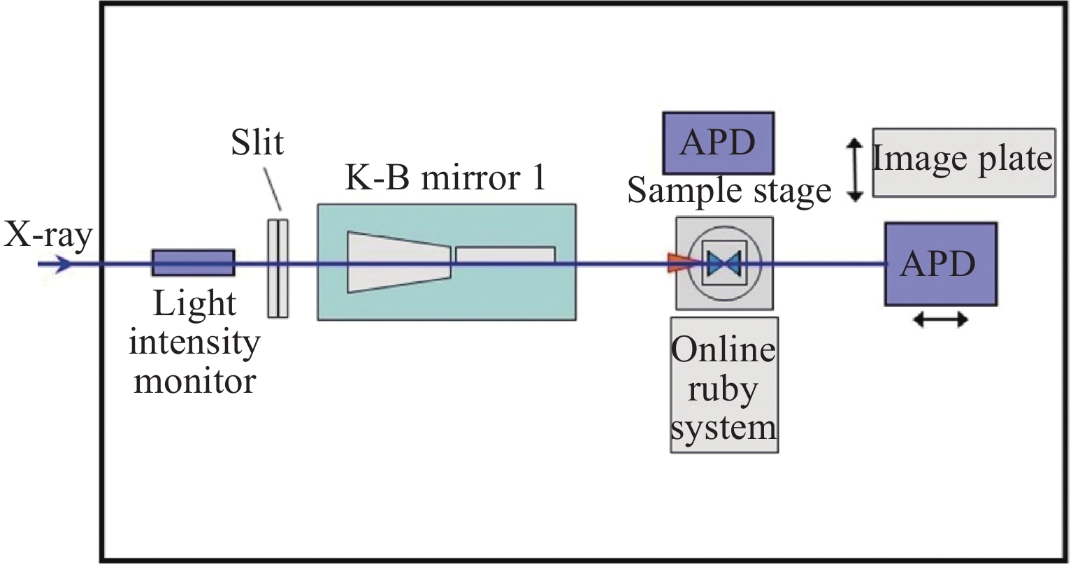
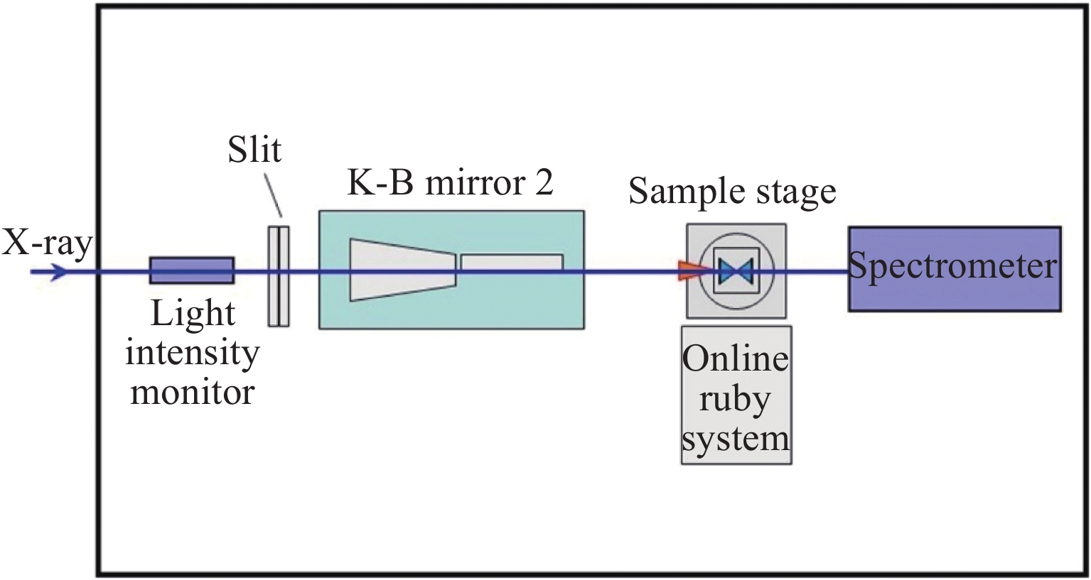
高压光束线实验站由两个棚屋组成,上游为微米XRD棚屋,下游为亚微米XRD棚屋。如图4(a)所示,微米XRD棚屋内有两个实验平台,分别为通用衍射平台(General purpose table,GPT)和激光加热专用平台(Laser heating table,LHT)。GPT除了完成常规的粉末、单晶、多晶以及径向XRD等实验,还可以开展原位(电阻加热)高/低温以及压力动态加载XRD等实验。LHT可以为用户提供红外波长(约为1 064 nm)的连续或脉冲激光加热实验条件,未来还可以提供CO2激光加热的实验条件。为满足实验设备及环境的稳定性要求,亚微米XRD实验平台(图4(b))搭建在实验站末端的专用亚微米XRD棚屋内,除了可以提供小光斑完成以XRD为基础的实验,未来还会逐步提供成像、相干等实验手段。
-
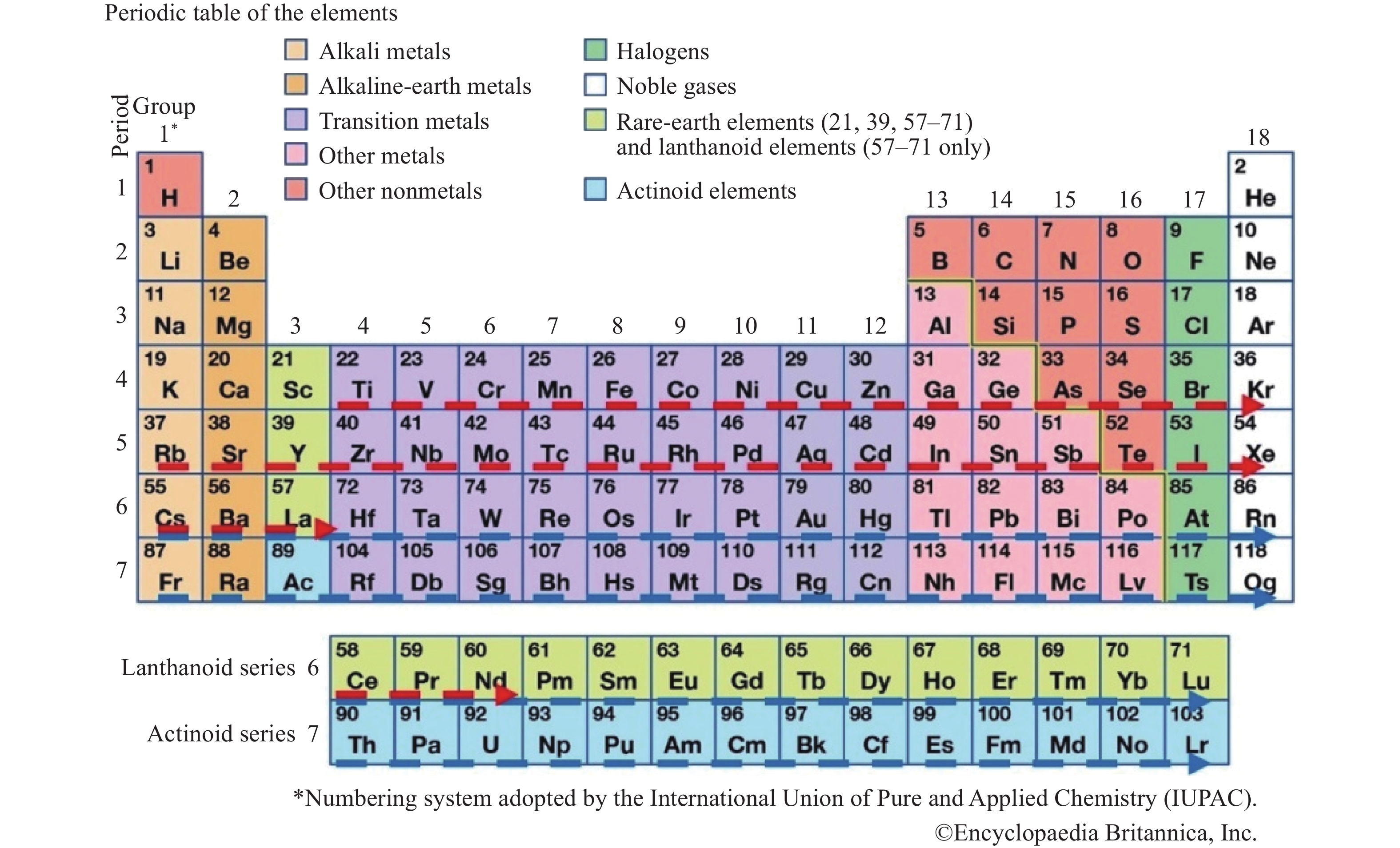
X射线吸收谱学(X-ray absorption spectroscopy,XAS)线站从HEPS储存环插入件ID46引出,是一条以XAS及相关衍生实验方法为主进行优化设计的高性能硬X射线光束线。XAS线站设计的基本目标是打造一个通用性强、能量覆盖范围大,同时具有高通量、高亮度的X射线吸收精细结构(X-ray absorption fine structure,XAFS)实验系统,为用户提供可靠、稳定、操作简便的XAS实验平台。该平台具备XAS的常规实验方法,具备时间分辨和空间分辨能力,能够探测微区、表面、界面结构。同时线站还将为用户提供多种原位样品环境,并结合XRF、XRD、FTIR、质谱等实验方法,提供更丰富的样品信息。XAS线站设计能量覆盖范围为4.8~45 keV,如图5所示,设计能量能够覆盖化学元素周期表中22号元素Ti到60号元素Nd的K边XAFS能量范围,以及55号元素Cs以后的L边能量范围。
在高压研究中,XAS可以对样品中特定元素的价态、局域结构(如键长、配位数、空间构型)进行表征,可以为高压相变路径的确认提供与XRD相互补充的结构信息。
-
XAS线站的设计能量范围为4.8 ~45 keV,最小聚焦光斑尺寸350 nm × 350 nm。工作能量在10 keV时,X射线大光斑(尺寸约为2 mm × 1.5 mm)的光通量在1013 ph/s水平,亚微米光斑(尺寸约为350 nm × 350 mm)的光通量在1012 ph/s。亚微米光斑由K-B镜组获得,其工作距离约为140 mm,可以满足常用DAC实验的空间要求。XAS线站的主要设计指标见表2。
-
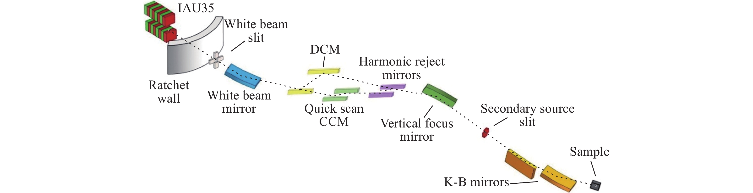
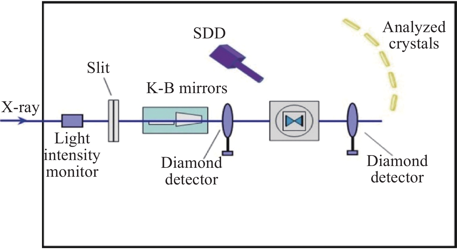
XAS线站光学元件布局如图6所示,X射线从储存环插入件ID46引出。真空外插入件(IAU)的磁周期长度为35 mm,总周期数141,设计最小gap值为11 mm。X射线通过白光压弯镜、双晶单色器(Double crystal monochromator,DCM)、快扫单色器(25毫秒每谱)、谐波镜组、垂直聚焦镜等主要光学元件后进入实验站,利用K-B聚焦镜组将X射线聚焦到样品点。实验站还配有后分析晶体及硅漂移探测器(SDD),主要用于高能量分辨吸收谱(HERFD-XAS)以及后续有待开发的低分辨发射谱(XES)。实验平台布局见图7。
-
硬X射线高分辨谱学(Hard X-ray high energy resolution spectroscopy,H2O)线站从储存环ID33插入件引出。该线站侧重于发展高分辨谱学实验方法,为用户提供具有高能量分辨率的核共振散射和X射线拉曼散射等实验手段,为凝聚态物理、化学、材料学、地学、环境等学科前沿研究提供有力工具[28]。线站建成后,将与世界上主要同步辐射光源的高分辨谱学站(如PETRAⅢ的P01线站、Spring-8的BL09XU线站、ESRF的ID18和ID20线站以及APS的3ID、20ID线站等)具有同等水平的实验和数据分析条件。
核共振散射[14]和X射线拉曼散射[15]能够与XRD等实验技术互补,在电子结构及晶格动力学方面给出独特的样品结构信息,尤其在高压研究领域具有举足轻重的作用。利用高压核共振散射方法可以获得关键热力学参数[29]、地球化学演化中的动力学行为[30]、地球深部的磁场信息[31]以及高压超导的物理机制[32-35]。X射线拉曼散射可以获得高压下轻元素C、N、O的吸收谱,是高压水[36]、二氧化碳[37]和二氧化硅[38]等谱学研究的利器,此外由于其具有非偶极跃迁的探测本领,还是开展稀土金属高压体积坍缩引起电子结构变化[39]等研究的重要工具。
-
HEPS光源有两种注入模式:一种是63个束团的高电荷模式,相邻束团间隔为72 ns;另一个是680束团的高亮度模式,束团间隔为7 ns。针对HEPS光源的注入束团模式,H2O线站将采用分时运行模式,以满足不同用户群体对高分辨谱学方法的需求。在高电荷模式下,可以实现57Fe的核共振散射实验,以及119Sn、151Eu等核同位素的向前散射实验和非弹性散射实验;在高亮度模式下,可以提供X射线拉曼实验条件。表3所示为H2O主要实验方法及相关技术指标。
-
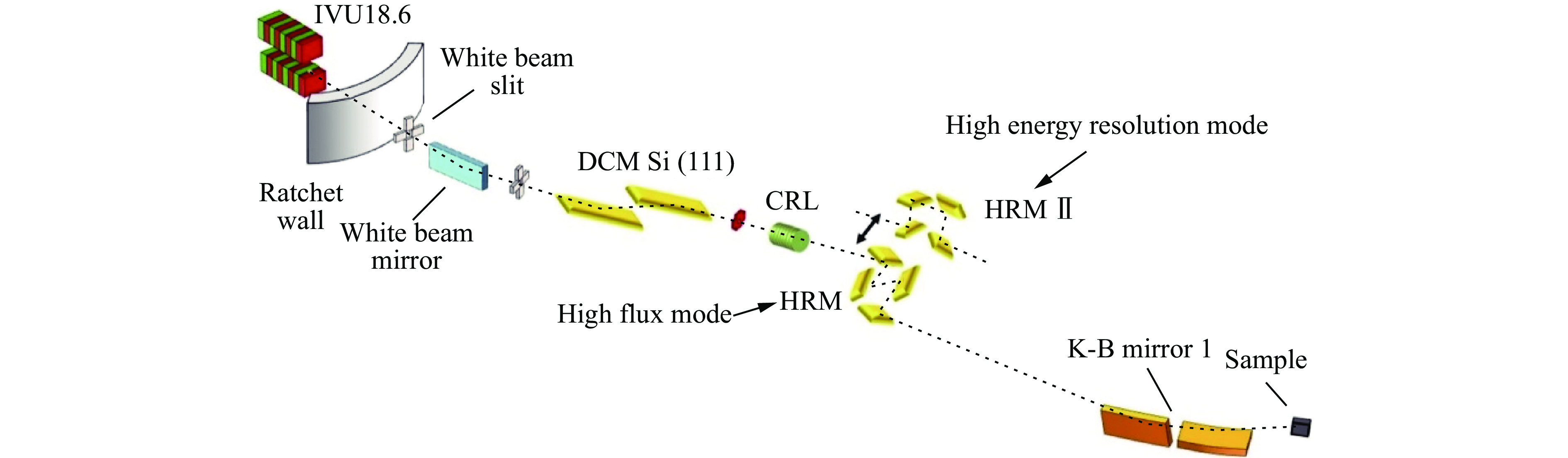
由于高分辨同步辐射光能量带宽极窄、散射截面小等原因,高分辨谱学线站多被称为“光子饥渴型”线站,因而需要获得极高的光子通量。为了实现高光子通量的设计目标,H2O线站选用真空内永磁波荡器插入件(IVU)作为光源。波荡器长4 m,周期长度为18.6 mm,最小磁间隙为5.2 mm,磁性材料为NdFeB。插入件基波能量覆盖7~17 keV,3次谐波可以覆盖21~51 keV,确保了57Fe核能级位于基波且保持较高的光子通量,同时确保Si(660)分析晶体背散射能量9.7 keV处的高通量,而3次谐波也将覆盖119Sn、151Eu、161Dy等常用同位素的核共振能级。
H2O线站光学元件布局如图8、图9所示。从插入件引出的X射线经前端区后被第1个水平偏转的白光反射镜反射,并吸收部分热量。白光反射镜下游的DCM将同步辐射光单色化(能量带宽10−4~10−5)。对于核共振散射模式(图8),经过DCM的X射线需要再进入高分辨单色器,进一步单色化至10−7量级以满足实验要求。DCM下游的CRL工作能量为14.4 keV,用于光路准直,以提高下游高分辨单色器和K-B镜对X射线的接收效率。X射线经K-B镜聚焦后可以获得微米尺寸的聚焦光斑,用于高压实验。在核共振散射实验中,部分研究需要极高的能量分辨率(如1 meV),但可以接受光通量减小,这时只需切换不同的高分辨单色器,并适当调整K-B镜和样品的位置即可。X射线拉曼散射实验模式光学布局如图9所示,X射线经Si(111)/Si(311) DCM单色化后通过K-B镜聚焦,到达拉曼散射谱仪完成实验。
如图10、图11所示,H2O实验站棚屋内有两个实验台。图10为核共振散射实验的光学平台,在该平台上K-B镜的工作距离可达1 m,并将搭载原位测压系统。核共振非弹性散射实验采用3个雪崩光电二极管APD探测器,分别位于DAC压砧的侧面;而时域穆斯堡尔谱的超精细结构谱学研究则采用透射模式,只采用单个APD探测器。该探测器移出后,可以将成像板探测器移入,采集样品的高压衍射信号。
X射线拉曼散射实验光学平台布局如图11所示,在一个纵向尺寸为4 m的拉曼散射谱仪上完成,图中省略了谱仪的详细设计图。可以看出,拉曼散射谱仪K-B镜的工作距离可达1.2 m,以确保拉曼散射可以采集到散射角大于150°时的高动量转移信息,这部分信息与电子的非偶极跃迁有关,从而可以提供丰富的电子结构信息。谱仪主要是由分析晶体和探测器组成,结构复杂,不再赘述。
-
X射线显微成像(Transmission X-ray microscopy,TXM)线站中,X射线从HEPS储存环插入件ID30引出,是开展基于波带片放大的全场成像实验方法为主的成像线站。TXM线站除可以开展全场吸收成像、泽尼克相衬成像外,还将重点发展近边谱学成像[40]实验方法,在三维形貌结构信息的基础上获取三维的元素化学价态等近边谱学信息及其演变过程信息。线站建成后,将和世界上主要同步辐射光源的全场纳米成像线站(APS的32-ID-C线站、NSLSⅡ的18-ID线站、SSRL的6-2c线站等)具有同等水平的实验及数据分析条件。
在高压研究中,X射线成像技术可以在诸多方面发挥作用:在物态方程测量中,X射线成像可以针对特定样品,尤其是非晶或无定形材料样品,为研究物质体积/密度对压力/温度的响应获取相应的信息[41-45];在相变演化及其动力学过程研究中,可以利用X射线成像技术得到密度、元素、价态等信息空间分布的特点,观测相变核的分布和相变过程[46-47];在地球科学相关机制的研究中,可以利用成像实验及相应的数据处理方法研究地幔中重要金属元素的输运机制[48-49]等。
-
TXM线站的设计能量范围为5~15 keV,最高空间分辨可达20 nm,成像视场大小根据具体空间分辨要求可在13~60 μm范围内变化,能量为8 keV时样品点的光通量在1012 ph/s水平。
以最高20 nm空间分辨成像时,受样品到波带片距离(波带片焦距约20 mm)的限制,成像用DAC的设计直径会受到一定的限制。此时,成像单元的半径须小于20 mm(15 mm为佳)。
-
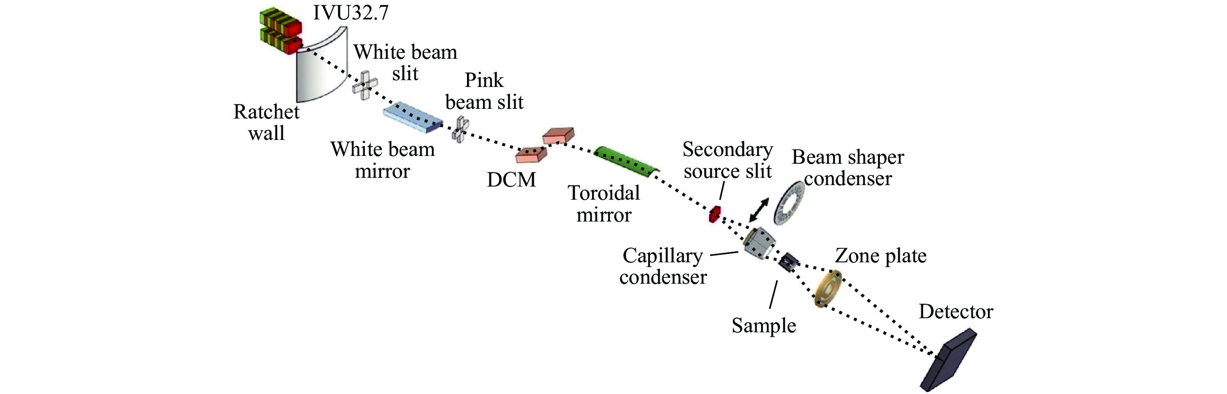
TXM线站光学布局如图12所示。同步辐射X射线从储存环插入件ID30引出,通过白光反射镜、DCM、超环面镜聚焦镜等主要光学元件后进入实验站。实验站利用椭球毛细管聚焦镜(Capillary condenser)或光栅型聚焦镜(Beam shaper condenser)将X射线聚焦到样品点并照明波带片,最后在探测器上获得样品的放大图像。
ID30插入件为真空外插入件(IAU),周期长度为32.7 mm,总周期数152,设计最小gap值为11 mm。因为波带片成像需要空心锥照明光束,所以结合HEPS储存环的低发射度的特点,TXM线站创新地采用了插入件红移照明设计[50],实现了空心锥照明的自然形成和插入件光子通量的最大化利用。
TXM线站设计使用Si(111)DCM选取需要能量的X射线,同时保证了适合近边谱学成像的能量分辨率。在DCM前设计了白光反射镜以缓解光束线的高热负载影响,白光反射镜采用Si和Rh两种反射层以达到谐波抑制的目的。在DCM后设计超环面镜(Toroidal mirror)作为初级聚焦,配合实验站的聚焦镜(Condenser)满足波带片成像所需的照明角度需求。
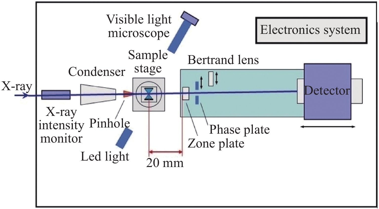
TXM线站实验平台布局如图13所示。实验站选择毛细管聚焦镜(Capillary condenser)满足高分辨(小成像视场)成像需求,以充分利用毛细管聚焦镜的高反射效率。同时,实验站还配备光栅型聚焦镜(Beam shaper condenser)以满足更多的成像视场需求。实验站的聚焦镜、波带片和探测器等都有沿光路方向移动的运动自由度,通过控制程序进行联动,从而实现不同X射线能量下整体成像系统的放大倍数一致性。成像时,样品的结构信息通过波带片放大后投射到探测器上,利用波带片和透镜耦合探测器的光学放大可以将纳米级的样品结构细节放大到探测器可以分辨的微米级,从而实现高分辨的全场成像。
-
HEPS建有专用或兼用的光束线站向用户开放,还配备了高压专用实验室,提供高压实验和样品准备所需的多种辅助设施。高压实验室包括样品准备实验室和光学实验室。样品准备实验室除了配备高倍显微镜、液氩装填等常规设备,还将配置脉冲激光打孔、手套箱、高压充气等配套设施,未来计划添置机械手、样品切割等设备;光学实验室将配置红宝石/拉曼测压、激光加热等设备。为了方便在非高压专用线站尽快建立适合高压实验所需的实验条件,还将准备便携的红宝石测压系统、激光加热系统,以及气膜加压等配套设备。如果有需要,压力动态加载、低温杜瓦、电阻加热等设备也可以从高压光束线站移动至其他线站共享使用。
2.1. 高压光束线站
2.1.1. HPB线站的主要技术指标
2.1.2. HPB线站设计
2.2. X射线吸收谱学线站
2.2.1. XAS线站主要技术指标
2.2.2. XAS线站设计
2.3. 硬X射线高分辨谱学线站
2.3.1. H2O线站的主要技术指标
2.3.2. H2O线站设计
2.4. X射线显微成像线站
2.4.1. TXM线站的主要技术指标
2.4.2. TXM线站设计
2.5. HEPS高压实验辅助设施
-
除了文中介绍的4条线站外,HEPS的一期建设计划还包括生物大分子微晶衍射线站、低维结构探针线站、高分辨纳米电子结构线站、硬X射线相干散射线站、粉光小角线站、工程材料线站、硬X射线纳米探针线站、硬X射线成像线站、结构动力学线站和通用环境谱学线站10条光束线站,以及1条主要用来进行同步辐射光学元件检测和实验技术发展的专用测试线站。在上述线站中,硬X射线相干散射线站、粉光小角线站、工程材料线站和硬X射线纳米探针线站等经过改造,也可以开展DAC高压相关实验研究,如XPCS、小角散射、PDF、超高压衍射等。限于篇幅,本文不再多作介绍。
作为公益性的科研设施,HEPS在考虑用户普遍需求的同时,也兼顾了一些特殊需求,致力于建设一个支撑多学科前沿研究和满足国家需求的重要实验设施。根据远景规划,HEPS未来要完成近百条光束线站的建设工作,其中将有6条专用的高压线站以及若干条可以开展高压实验的兼用线站。专用的高压线站中,除了目前已经在建的XRD线站,还包括另外两条衍射或散射线站和一条谱学线站,以及两条混合方法线站。为了更好地完成高压相关线站的规划,欢迎广大用户对后续的建设方案和方法选择提出意见和建议。HEPS光束线站建设尤其是高压线站群的建设,将为高压相关领域研究提供重要的支持手段。
感谢HEPS项目组光束线站部中高压光束线站系统、X射线吸收谱学线站系统、硬X射线高分辨谱学线站系统和X射线显微成像线站系统工作人员的辛勤付出,感谢线站部光学系统、光机系统、控制系统和通用机械系统等对设计工作的支持与配合。感谢上海光源、合肥光源、APS、SPring-8、ESRF、PETRA Ⅲ和NSLS Ⅱ等光源相关线站科学家的支持和帮助,感谢国内外高校及科研院所用户代表对项目建设初期的建议和支持。感谢HEPS国际顾问委员会对线站布局定位的意见和提议。