-
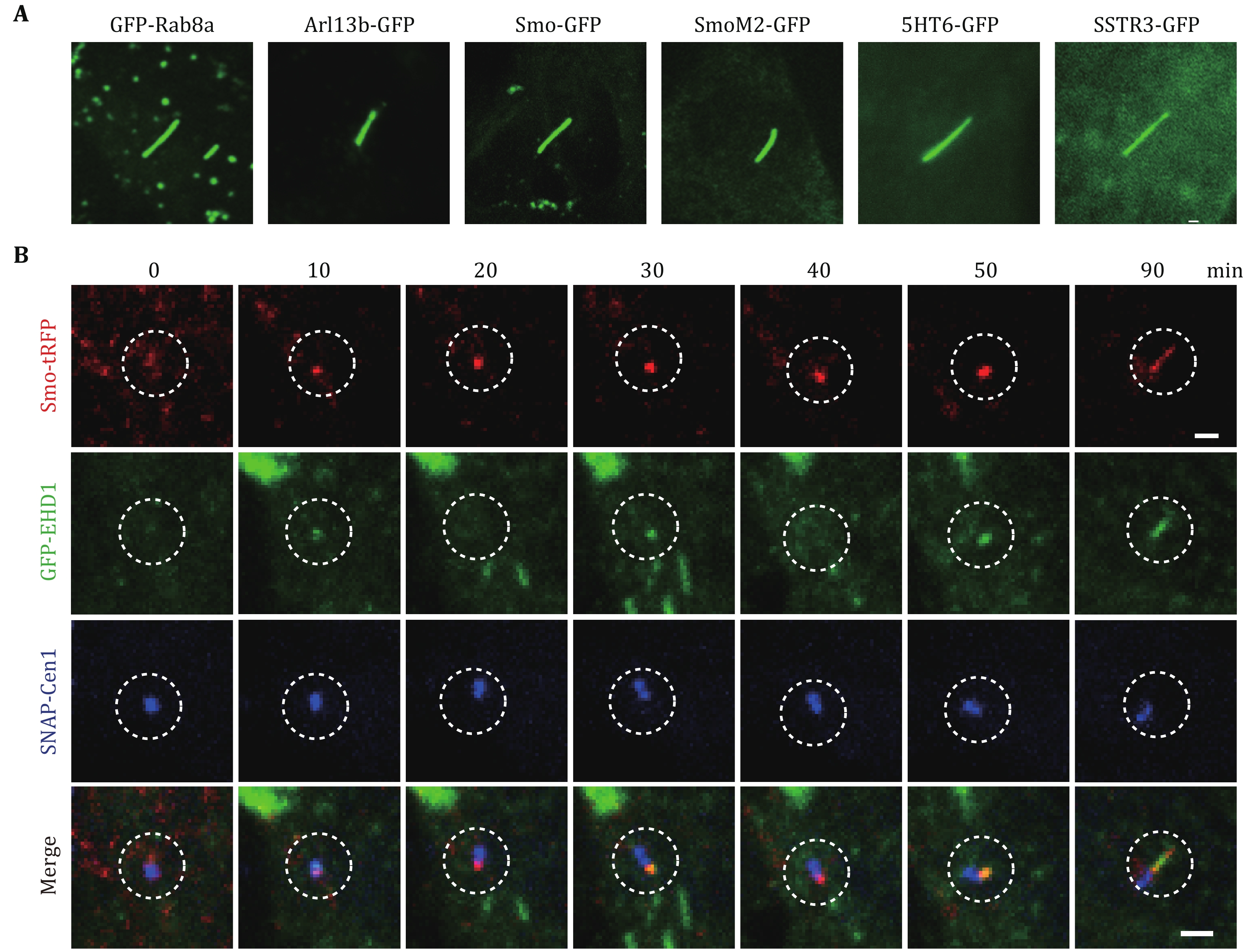
Figure 1. Ciliary markers and ciliogenesis in RPE-1 cells. A GFP-tagged Rab8a, Arl13b, SMO, SmoM2, 5HT6 and SSTR3 localize to primary cilia in RPE-1 cells. Images are captured in live cells with epifluorescence microscopy. Scale bar, 2 µm. B Ciliary membrane assembly was imaged using spinning disk confocal microscopy. SMO-tRFP and SNAP-Centrin1 mark the ciliary membrane and centrosome, respectively. SNAP-centrin1 was stained with SNAP-Cell 647-SiR for 30 min before imaging. Live-cell imaging demonstrates the dynamics of GFP-EHD1 localization in the developing ciliary membrane. Image series show the ciliary membrane assembly process from vesicle docking to cilia elongation. Images were captured every 10 min. Scale bar, 2 µm
-
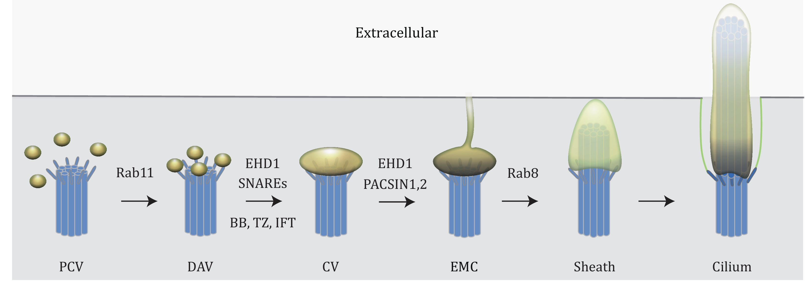
Figure 2. Sequential steps of primary cilium assembly revealed by live-cell imaging. Model of intracellular ciliogenesis. At the early stage of ciliogenesis, pre-ciliary vesicles (PCVs) are transported to the mother centriole through a Rab11-mediated membrane trafficking pathway. Some of these vesicles dock onto the mother centriole's distal appendages and become distal appendage vesicles (DAVs). EDH1 and SNARE proteins promote the reshaping and fusion of DAVs to form a larger ciliary vesicle (CV). The reorganization of membrane vesicles from DAV to CV is a prerequisite step for the basal body (BB) and transition zone (TZ) formation and IFT proteins recruitment. Remarkably, EHD1 and PACSIN1/2 assemble membrane tubules from the developing intracellular cilium that can connect to the plasma membrane and create an extracellular membrane channel (EMC) to the outside of the cell. Rab8 promotes ciliary sheath growth from the CV and the intracellular ciliary sheath subsequently fuses with the plasma membrane and the exposing the developing cilium is exposed to the extracellular environment. The sequential assembly of CV, BB, TZ and IFT was revealed by live-cell imaging (Lu et al. 2015a). The application of live-cell imaging shed new light on mechanisms of ciliogenesis regulation
-

Figure 3. Comparison of epifluorescence, spinning disk confocal, image deconvolution and TIRF microscopies in primary cilia imaging. RPE-1 cells expressing ciliary pocket marker GFP-EHD1 and ciliary membrane marker Rab8a-tRFP were imaged with different microscopy modalities using a Marianas inverted microscope system (Intelligent Imaging Innovations). Note the differences in the signal-to-noise ratio of microscopy techniques. Scale bar, 5 µm
-

Figure 4. Dynamics of GFP-EHD1 and tRFP-Rab8a at the ciliary pocket and ciliary membrane by TIRFM. Cilium in RPE-1 cells expressing GFP-EHD1 and tRFP-Rab8 was imaged with TIRFM. Note that GFP-EHD1 and tRFP-Rab8a localized on dynamic membrane tubules at the ciliary pocket membrane. The figure showing images captured every 30 sec. The signal intensity of tRFP-Rab8a was inverted and enhanced to demonstrate the presence of Rab8a in the membrane tubules at the base of cilia. Scale bar, 5 µm
Figure
4 ,Table
1 个